Bone Health after Spinal Cord Injury
Spinal cord injury (SCI) causes paralysis and as a result, the mechanical stimulation to the skeleton normally provided by muscle activity and weight bearing is severely attenuated. Bone loss is a prominent feature of the immediate post-injury period, however fracture rate does not increase significantly until 5-10 years post-injury. These features make individuals with SCI a model population in which bone adaptive response due to lack of mechanical stimulus may be investigated. Additionally, because the SCI population is at high risk for fractures (similar to that of osteoporotic women), individuals could benefit from interventions that reduce loss of bone mass and strength. Anti-resorptive therapy, a key intervention for osteoporosis, cannot fully ameliorate this problem. Novel therapies and a more complete understanding of the structural changes that occur within the bone of SCI patients would benefit this population. Mechanical vibration is one novel therapy that has shown promise for improving bone health. Teriparatide is anabolic to bone and is currently approved for clinical use in some populations.
Sub-projects related to spinal cord injury:
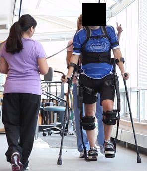
A patient walking with the assistance of the Ekso
Skeletal and Clinical Effects of Exoskeleton-Assisted Walking Therapy in Individuals with Spinal Cord Injury
This biomechanics study is a collaborative project taking place at Spaulding Rehabilitation Hospital in Boston with colleagues Leslie Morse, DO, and Paolo Bonato PhD (Troy/Bonato/Morse co-PIs; funding: Department of Defense). Although exoskeletons assist individuals in reproducing gait kinematics, the actual forces occurring within the musculoskeletal system are dissimilar to those occurring during natural walking. Given that fractures have occurred during assisted gait retraining, understanding these forces and torques in the context of the underlying bone strength is a critical safety issue for broad implementation of these devices. Furthermore, the degree to which exoskeleton-assisted ambulation may increase bone and muscle mass in individuals with SCI is not known, although evidence suggests that the forces generated through assisted gait may be sufficient to stimulate improvements to both tissues. The purpose of the project is to quantify the forces and moments placed on the skeletons of individuals with SCI while they walk in a commercially-available exoskeleton, the Ekso.
Effects of Ekso-Assisted Walking Therapy on Bone Health and Quality of Life: A Randomized Clinical Trial
This clinical trial will examine the effects of a 6-month exoskeleton walking intervention on bone structure and strength, and quality of life in people with spinal cord injury. PI Leslie Morse will coordinate therapy, and the MBL will be working on bone-related outcome measures and data analysis. Information about the clinical trial can be found at ClinicalTrials.gov trial #NCT02533713.
Mechanical consequence of bone loss in acute spinal cord injury

Finite element model of a subject’s proximal femur, simulating a fall to the side. These models were generated from CT data and are one example of how subject-specific estimations of fracture strength can be made (Edwards et al., JBMR 2014)
Spinal cord injury is characterized by marked bone loss at sublesional regions. The bone loss occurs rapidly, with the greatest reductions being observed at the knee. Within the first 6-18 months of spinal cord injury, approximately 50% of the initial bone mass has been resorbed at the distal femur and proximal tibia. The biomechanical significance of these changes is not fully understood and we are conducting a study to quantify changes in whole bone stiffness resulting from acute (<1 year) spinal cord injury. Former lab member W. Brent Edwards developed subject-specific finite element modeling methods to quantify changes in torsional and compressive stiffness of the distal femur and proximal tibia. This work has shown that over a 3 month period post acute spinal cord injury, relative reductions in whole bone stiffness are approximately 1.5 to 2 times greater than relative reductions in bone mineral. These data suggest that simple measures of bone mineral may underestimate the effect that spinal cord injury has on the mechanical integrity of bone.
Effect of Teriparatide, vibration, and the combination, on bone mass and bone architecture in chronic spinal cord injury
This trial was recently completed in collaboration with Thomas J. Schnizter, MD, PhD (Department of Defense, PI Schnitzer) and W. Brent Edwards. The purpose of the trial is to determine the clinical efficacy of teriparatide, a bone anabolic agent, mechanical vibration, also shown to be anabolic to bone in some situations, and a combination therapy. Our lab developed quantitative CT metrics that will be used to evaluate the success or failure of the interventions: three-dimensional changes to bone mass, density, and shape that occur over time. The trial period was recently completed and data analysis is ongoing.