Measurement of bone deformity in Rheumatoid and Psoriatic Arthritis
Rheumatoid arthritis (RA) and Psoriatic Arthritis (PsA) are two types of chronic inflammatory arthritis that result in progressive disability and joint degeneration. RA results in abnormalities in bone metabolism, including the generation of focal bone erosion sites within joints and patients with PsA develop pathologic bone formations (osteophytes). The joints of the hand are commonly affected in both disorders, with pain, swelling, and skeletal deformity being a primary cause of disability.
High-resolution computed tomography (HR-pQCT) has been used to quantify the presence and size of focal erosion sites or osteophytes in individuals with RA and PsA. However, quantification of erosion and osteophyte size depends on knowledge of the original bone surface, which has presented methodolo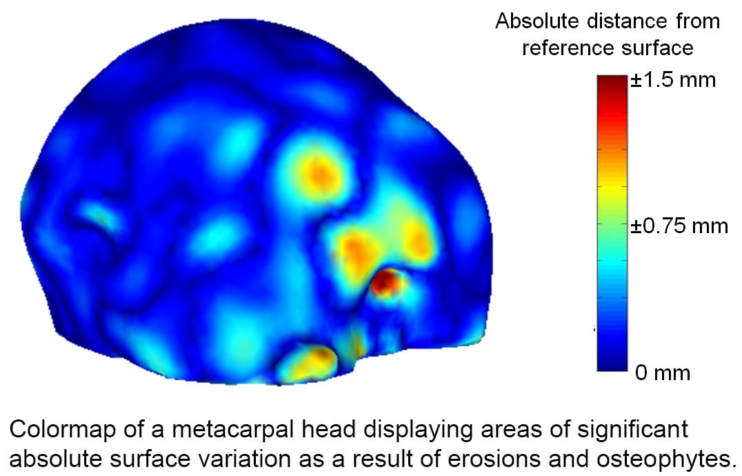 gical challenges.
An analysis method called statistical shape modeling is used to quantify bone surface morphology, which can objectively and reproducibly quantify statistical deviations from “normal”. The objective of this study is to develop novel quantitative measures that define joint deformity in RA and PsA, and associate these measures with clinical parameters and with biomarkers of disease state. The figure on the left illustrates how osteophytes and erosions can be measured objectively in diseased bone compared to a reference surface.
This project is in collaboration with Dr. Ellen Gravallese at the University of Massachusetts Medical Center, and is funded by a WPI/UMass Collaborative Seed Grant.